
Step by step on the way through 9 months
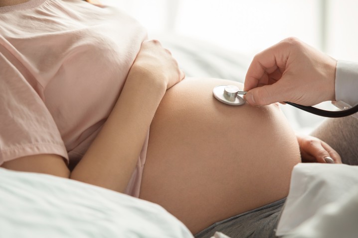
Adequate monitoring of pregnancy can diagnose possible problems with the fetus in time, but also warn of possible health problems that may develop in the mother. That is why regular visits to the doctor are of great importance.
Pregnancy and motherhood are special for every woman. From the moment a woman finds out she is pregnant until the first cry of the baby, the pregnancy period is full of surprises, changes and adjustments to future motherhood.
That is why monitoring pregnancy is necessary for the safe growth and development of the child, but also for taking care of the mother’s health.
Adequate monitoring of pregnancy can diagnose possible problems with the fetus in time, but also warn of possible health problems that may develop in the mother. That is why regular visits to the doctor are of great importance.
Going for check-ups is an unavoidable part of every pregnancy.
From the first check-up to going to the hospital just before giving birth, monitoring your pregnancy is extremely important because of the observation of you and your baby. Regular check-ups closely monitor any changes in the growth and development of the fetus and prevent the occurrence of possible complications.
Diagnostic methods vary depending on the period of pregnancy you are in. Regardless of whether it is the first or third trimester, some examinations can be repeated throughout the entire period of pregnancy, while others can only be done at a certain stage of pregnancy.
Some of the examinations that are carried out several times are certainly ultrasound and laboratory and vaginal tests through which we record the growth and development of the fetus, but also take care of the mother’s health and the prevention of possible vaginal infections.
In the first stage of pregnancy, the fertilized egg descends into the uterus to continue its development. After the 6th week, a primitive muscle is formed that will later develop into the heart, and thus the process of blood circulation through the embryo begins.
By the end of the 12th week, the fetus begins to develop reproductive organs, fingers and toes, nails appear, and facial features are formed.
In the first trimester, the fetus will be about 6 cm long, which is why you will not be able to feel it yet.
The beginning of your 9-month journey begins with going to the first check-up where the gynecologist confirms your pregnancy and determines the exact date of delivery, as well as appointments for future check-ups and pregnancy monitoring.
Before carrying out any tests, our staff will first speak to you. During the consultation, we will go through the topics of your menstrual cycle, general health, but also provide certain guidelines that you should follow in the coming period.
During the conversation, you will also touch on the topics of previous pregnancies (if any), miscarriages and symptoms you may have experienced (morning sickness or increased discharge).
After the consultation, the first examination is carried out, which includes:
Speculum and bimanual examination examine your genitals in detail, and based on indicators such as blood circulation and secretion of discharge, pregnancy is confirmed.
Ultrasound can be performed in the 6th week of pregnancy, and due to the size of the embryo that cannot be detected by the standard method, we use transvaginal ultrasound.
By taking swabs and conducting laboratory tests, we gain insight into the presence of possible vaginal infections, but also into the complete blood count (CBC).
Although ultrasound in the early phase (6th week) is not mandatory and depends on the gynecologist’s assessment, the one in between Weeks 10 and 14 It is certainly one of the most important in monitoring pregnancy.
This ultrasound can monitor the vital functions of the fetus and detect possible developmental anomalies. Indicators of such changes can be observed by observing the width of the nuchal fold of the fetus and the presence of the nasal bone.
These abnormalities may indicate fetal chromosomal anomalies and the detection of Down syndrome or some other chromosomal pathies (Turner sy., Edwards sy., Patau sy., etc.).
The second trimester of pregnancy is the period when pregnancy becomes visible. The uterus comes out of the small pelvis, which causes the growth of the abdomen, breast enlargement, and weight gain is also recorded.
In this period of pregnancy, the fetus is now easily recognizable, with visibly formed parts of the body. Although the fetus is only 23 cm long, it can grab with its hands, kick with its legs and make rolls.
Now he reacts to sounds and touch, develops swallowing and breathing reflexes, and hair begins to grow on his head. The movement of the fetus in the womb can be felt.
For this stage of pregnancy, regular gynecological examinations are important, of which ultrasound examinations stand out. Examinations are carried out during the second trimester in the 15th, 20th and 25th week of pregnancy, which records changes in growth, heart function and movements inside the uterus.
In addition to conducting ultrasound (between the 18th and 22nd week of pregnancy), other diagnostic examinations are carried out at this stage of pregnancy:
Since the fetus is more developed at this stage of pregnancy, the ultrasound that is performed during this period is not only used to observe the growth of the fetus, but is also used to identify possible anomalies that could not be detected before. This type of ultrasound is also called an anomaly scan.
During this ultrasound, the gynecologist examines all organs and organ systems, the placenta, the umbilical cord and the amniotic fluid.
By taking a sample of amniotic fluid , chromosomal disorders that were not visible in the first trimester can be diagnosed. This type of examination is called amniocentesis. In addition to amniotic fluid, the placenta (choriocintesis) can also be used for additional tests.
The advantage of these examinations is reliability, but due to the complexity and possible complications of these invasive methods, there is an increased risk of spontaneous abortion.
The last trimester of pregnancy is now clearly recognizable, and the baby is gradually preparing to go out. The baby descends into the birth canal after the 36th week and turns with the head down. The body’s functions are fully developed, and the baby is about 46-56 cm long.
Due to weight gain and growth, the baby is now putting more and more pressure on the mother’s body, which is why back pain, digestive problems, pressure on the bladder and breathing disorders are possible. In the last stage of pregnancy, many mothers complain of insomnia and the more frequent occurrence of false labor.
As for examinations, it should be noted that after the 35th week of pregnancy, they are performed in the hospital where pregnant women plan to give birth.
At this stage of pregnancy, examinations such as measuring the weight of the pregnant woman, blood pressure and ultrasound are carried out again, and taking a vaginal and rectal swab for BHSB and CTG recording are particularly noteworthy.
BHSB, also known as group B hemolytic streptococcus, is a bacterium that can occur in pregnant women. In case the bacterium is present during childbirth, it can pass from mother to baby.
Contact with this bacterium can cause diseases such as sepsis, pneumonia and meningitis in the baby. To prevent infection with this bacterium, a swab is taken from the vagina through which the possible presence of the bacterium can be determined.
In case of positive results, doctors decide on antibiotic therapy, most often penicillin. It should be noted that antibiotics for the treatment of this bacterium are administered exclusively intravenously and during childbirth. IT IS NOT NECESSARY TO TAKE THERAPY BEFORE CHILDBIRTH!
If the due date is exceeded, it is necessary, if the genital finding is favorable for this, to perform an amnioscopy.
Amnioscopy is an examination with which, if the cervical canal is passable, an attempt is made to see the color of the amniotic fluid (clear, milky, green, bloody, etc.).
Recovery after childbirth is considered almost as important as monitoring your pregnancy. Gynecological examinations are often done in the 6th week after delivery, and it is possible to leave earlier in case there were certain complications.
Gynecological examinations observe how your body recovers after childbirth. During the examination, we pay special attention to the size of the uterus, which should gradually shrink and return to its original state within two months.
If childbirth was carried out by cesarean section, we observe the rate of wound healing and possible indicators of infection.
In addition to causing physical exhaustion, the period of pregnancy could also leave a mark on your mental health.
In a conversation with our team of experts, we will go over topics such as postpartum depression, a common phenomenon that occurs after childbirth, and can be recognized by listlessness, anxiety and general depression of the mother.
This phenomenon, also known as baby blues, occurs due to a sudden change in hormone secretion that occurs 2-3 days after birth. A drop in estrogen, progesterone and prolactin levels can cause mood swings and depressed maternal behavior.
This phenomenon is certainly not harmless, and that is why we provide the possibility of an open dialogue during which you can tell us everything that is bothering you.
At the Lohuis Filipović Polyclinic, we have gathered a team of experts in the field of gynecology and endocrinology who will closely monitor each phase of your pregnancy and allow the 9-month period to pass without any difficulties.
Thanks to modern technology, we conduct a series of diagnostic examinations that give us quick and precise results, while warning of possible complications.
The health of you and your child is our priority, and by adequately monitoring the pregnancy, we try to keep it unchanged.
Make an appointment today by calling +3851 2444 646, or by filling out our online form.
The main goal of LF Polyclinic is to improve the quality of life and health of our clients by providing them with top-notch healthcare services.