
Find out all about the most important ultrasound in pregnancy
Ultrasound examinations in pregnancy are extremely important in monitoring pregnancy and early detection of possible anomalies and pathological conditions in pregnancy.
Ultrasound examinations in pregnancy are extremely important in monitoring pregnancy and early detection of possible anomalies and pathological conditions in pregnancy.
Ultrasound is a completely harmless, painless examination for which there is no alternative in monitoring a pregnant woman and a baby.
In addition to its medical significance, an ultrasound examination during pregnancy is also the first contact of future parents with their baby. For expectant mothers and fathers, it can be an intimate and touching experience in which they can see their baby’s movements and find out its gender.
An anomaly scan is a detailed ultrasound scan that is usually performed between the 19th and 22nd week of pregnancy, and allows the gynecologist to examine the organs and organ systems of the fetus.
Anomaly scan is used to try to find out the sex of the fetus, possible developmental anomalies, placental site and its pathological changes, amniotic fluid collection, and a whole range of abnormalities associated with pregnancy.
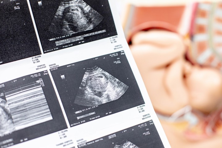
Ultrasound examinations are imaging diagnostic methods that allow ultrasound waves to visualize the baby in the mother’s body.
In pregnancy, ultrasound is used to safely confirm pregnancy, detect the number and sex of the fetus, monitor the development of the baby, and detect possible complications.
An anomaly scan takes longer than a classic gynecological ultrasound. During the examination, the gynecologist tries to show all the organs and organ systems of the child by examining the correctness of their development, umbilical cord, placenta and amniotic fluid.
As an integral part of the examination, cervicometry is also performed, i.e. measuring the length of the cervix and examining the internal uterine orifice.
Anomaly scan is part of routine ultrasound examinations, and is recommended for all pregnant women. The fetus is observed in the period of development, which is the ideal time to assess the appearance of the organ and detect any deviations from normal.
This test is extremely important because you will get to know your baby in detail and, if desired, find out the sex of the baby. You are free to take your partner or family member to the anomaly scan to make the experience even more special.
Ultrasound examinations are performed routinely in modern medicine. They represent one of the safest medical tests and have not proven to be harmful to the mother or child through decades of use.
According to the recommendation of the European Association of Perinatal Medicine, it is recommended to do a minimum of 3 ultrasound examinations during pregnancy. In the case of a high-risk pregnancy, the gynecologist may also recommend more frequent ultrasound examinations.
If the patient is not sure about the date of the last menstrual period and the time of conception, an early ultrasound examination can reliably determine the actual duration of pregnancy. The first ultrasound examination also determines the site of pregnancy and the number of fetuses.
The first regular ultrasound examination should be done between weeks 7 and 10 Pregnancy. Up to the 14th week of pregnancy, it is recommended to perform an ultrasound examination vaginally for a better view of the fetus and possible early anomalies and pathological changes in pregnancy.
So, at the first ultrasound examination, you will find out what your approximate due date is and whether the pregnancy is monotonous or more fertile, or how many children you are expecting. If they are twins, it is possible to find out at this examination whether they are monozygotic or dizygotic.
An ultrasound scan in the 15th week of pregnancy is sometimes called a mini anomaly scan. During this examination, the heartbeat is listened to, the available organs and organ systems are examined, the presence of “soft markers” for aneuploidies is examined, and the possible pathology of the placenta is also determined.
A mini anomaly scan cannot confirm the existence of diseases, but only indicates the possible existence of a higher risk for the existence of them. For accurate diagnostics, it is necessary to combine it with other diagnostic methods.
The examination is performed abdominally, i.e. with an ultrasound probe over the mother’s abdomen.
Anomaly scan is performed in the mentioned period of pregnancy because during this period the relationship between the amount of amniotic fluid and the size of the baby allows for a detailed examination of anatomy, assessment of movement dynamics and noticing possible anomalies in the development of the fetus and placenta.
Anomaly scan is an early screening method for chromosomal anomalies.
Although most babies are healthy, about 2-3 out of every 100 babies may have a perceived anomaly or condition that would require specialized care and more frequent supervision.
This ultrasound examination can also detect some minor deviations that do not pose a risk to the baby and do not require treatment.
During the anomaly scan, in addition to observing the anatomy of organs and organ systems, the measurement of fetal biometry is performed, which includes the diameter of the head, the circumference of the head, the circumference of the abdomen and the length of the femur.
In addition to the above, you can also find out the sex of the child at the examination. The same should be emphasized to the gynecologist in advance, that is, if you do not want to find out the gender, be sure to communicate it at the beginning of the examination.
It is important to note that an anomaly scan cannot detect all the anomalies that a child can be born with, but it can rule out a large number of them.
The value of the anomaly scan is in the early detection of the risk for the development of chromosomal anomalies, and possible morphological defects of the fetus that require more detailed care during pregnancy. This enables adequate preparation of the medical team in order to start treatment on time, i.e. to provide the newborn with all the necessary care and the best possible outcome of treatment.
Although the purpose of this ultrasound examination is to detect anomalies, it is necessary to emphasize that most of the results of the anomaly scan are a neat finding.
For most future parents, an anomaly scan is a sure confirmation that their baby is developing properly and that the pregnancy is proceeding normally.
An anomaly scan takes an average of 30 to 45 minutes. Special preparation for the examination is not required.
We recommend that you come to our Gynecology Clinic in comfortable clothes. You can consume food and drink before the examination without restrictions, but for examinations in the early stages of pregnancy, it is necessary to have an empty bladder.
The anomaly scan is performed on the abdomen. Before the ultrasound examination, the gynecologist will apply a gel to your skin that allows you to get a quality image of the baby.
For women for whom this is their first pregnancy, the anomaly scan represents a truly special moment. For the first time, you are introduced to your baby and the reality of future motherhood.
We definitely recommend that you bring your partner or family member with whom you want to share this unique experience.
The gynecology clinic of the Lohuis Filipović Polyclinic provides medical care using the most modern methods of diagnosis and treatment during pregnancy.
We especially nurture collaborative and teamwork that unites experience, knowledge and cutting-edge technology to respond to the specific needs of each patient.
Our goal is successful diagnosis, treatment, patient monitoring, and positive and long-term results.
The main goal of LF Polyclinic is to improve the quality of life and health of our clients by providing them with top-notch healthcare services.